Primary Retroperitoneal Mucinous Cystadenoma: A Rare Retroperitoneal Tumour
Primary Retroperitoneal Mucinous Cystadenoma (PRMC) is an exceptionally rare benign cystic tumour that arises within the retroperitoneum, the space behind the abdominal cavity. Unlike ovarian mucinous cystadenomas, PRMC has no direct connection to the ovaries or gastrointestinal tract, making its origin a subject of ongoing research.
Fewer than 200 cases have been reported worldwide, with most occurring in women between the third and sixth decades of life. Because of its rarity and non-specific symptoms, PRMC is often mistaken for ovarian cysts, mesenteric cysts, pancreatic cysts, or adrenal masses before surgery.
Complete surgical removal is both the definitive treatment and the only way to establish a definitive diagnosis.
Patient Presentation
A 35 years old lady presented with right iliac fossa pain and fullness for 3months.
- Normal and regular menses.
- She was nulliparous.
- Normal Bowel habit.
- No family history of malignancy.
Examination revealed anon mobile mass at right iliac fossa. Her laboratories were normal.
Diagnostic Evaluation
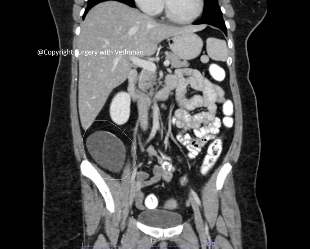
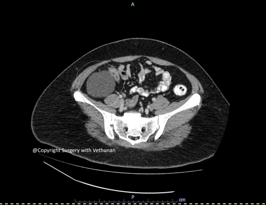
Contrast-Enhanced CT Scan
- Large, well-circumscribed thin walled cystic mass at right retroperitoneum region
- Homogeneous fluid contents
- Absence of obvious invasion into surrounding organs
- Displacement of right colon medially.
- Separate from kidneys, ureter and duodenum.
Surgical Management
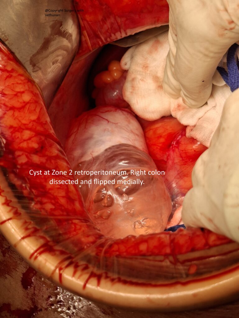
Excision was done through a midline technique rather than a laparoscopy as to prevent rupture of the thinned wall cyst.
The key surgical principles followed include:
- Complete en bloc removal
- Avoidance of cyst rupture
- Preservation of adjacent organs (right colon, ureter, kidney and duodenum)
- Retrieval of the specimen without spillage
Avoiding rupture is important because mucin spillage may complicate histopathological interpretation and, in malignant lesions, theoretically increase the risk of peritoneal dissemination.
Histopathological Examination
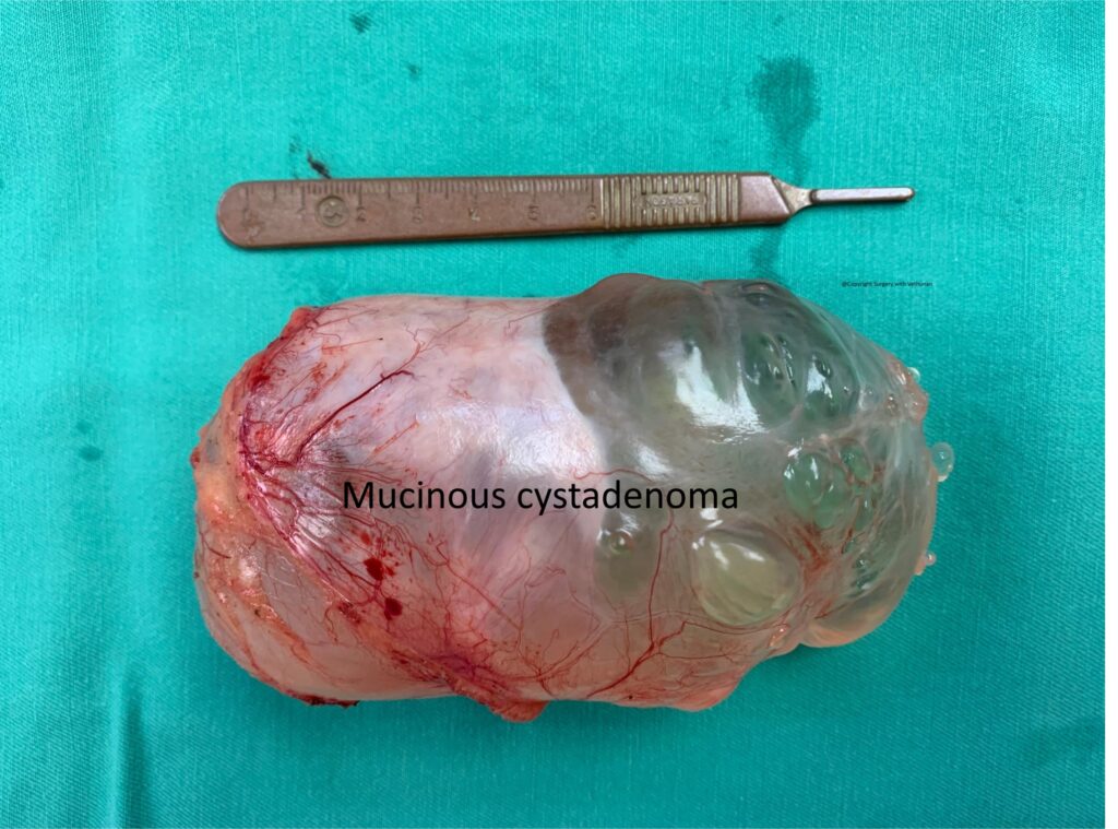
Final histopathological examination revealed:
Benign cystadenomas
- Mucin-producing columnar epithelium
- No stromal invasion
- No significant cellular atypia
Understanding Primary Retroperitoneal Mucinous Cystadenoma
The exact origin of PRMC remains uncertain.
Several theories have been proposed:
Coelomic Metaplasia Theory
The most widely accepted hypothesis suggests that remnants of embryonic coelomic epithelium undergo mucinous transformation over time.
Ectopic Ovarian Tissue Theory
Some lesions may arise from ectopic ovarian tissue located within the retroperitoneum.
Müllerian Remnant Theory
Embryological Müllerian remnants may differentiate into mucin-producing epithelium.
Despite these theories, the precise pathogenesis remains unclear due to the rarity of the disease.
Differential Diagnosis
Retroperitoneal cystic masses include:
- Mesenteric cyst
- Lymphangioma
- Pancreatic pseudocyst
- Adrenal cyst
- Renal cyst
- Cystic teratoma
- Ovarian cystic neoplasm
- Duplication cyst
- Hydatid cyst
Accurate diagnosis usually requires a combination of imaging, intra-operative findings, and histopathological examination.
Prognosis
The prognosis for benign Primary Retroperitoneal Mucinous Cystadenoma is excellent following complete surgical excision.
Key points include:
- Very low recurrence after complete removal
- Excellent long-term survival
- No chemotherapy or radiotherapy required for benign disease
- Periodic imaging follow-up may be recommended for selected patients
Borderline and malignant variants require closer surveillance and multidisciplinary management.
Why This Case Is Unique
Primary Retroperitoneal Mucinous Cystadenoma is one of the rarest cystic tumours encountered in general surgery.
This case highlights:
- An exceptionally rare retroperitoneal tumour
- The diagnostic challenge posed by large retroperitoneal cystic masses
- The importance of advanced cross-sectional imaging for surgical planning
- Successful complete surgical excision while preserving surrounding organs
- The essential role of histopathological examination in confirming the diagnosis
Key Takeaways
- Primary Retroperitoneal Mucinous Cystadenoma is an exceptionally rare benign retroperitoneal tumour.
- Symptoms are often vague, and many lesions are discovered incidentally.
- CT and MRI are essential for preoperative assessment but cannot reliably distinguish benign from malignant lesions.
- Complete surgical excision without cyst rupture remains the gold standard treatment.
- Histopathological examination is required to establish the definitive diagnosis and exclude borderline or malignant disease.
- Following complete excision, patients with benign lesions generally have an excellent prognosis.
Consent for online publishing of image and case obtained from patient