
Interstitial (Interparietal) Hernia: A Rare Hernia Variant Successfully Managed with Laparoscopic TAPP Repair – A Two-Case Series
Interstitial hernia, also known as an interparietal hernia, is an exceptionally rare variant of abdominal wall hernia in which the hernia sac lies between the layers of the abdominal wall musculature, rather than following the normal course through the inguinal canal.
It accounts for less than 1% of all abdominal wall hernias and is frequently overlooked because of its atypical clinical presentation.
Unlike conventional inguinal hernias, patients may present with diffuse abdominal wall swelling, chronic groin pain, lumbar swelling, or an ill-defined mass without a classical inguinal bulge. Consequently, diagnosis can be challenging, and many cases are only recognised during surgery.
The development of laparoscopic Transabdominal Preperitoneal (TAPP) repair has revolutionised the management of these uncommon hernias by allowing complete visualisation of the myopectineal orifice, identification of occult defects, and definitive mesh repair.
This case series highlights two patients with distinctly different presentations of interstitial hernia, both successfully diagnosed and treated using a laparoscopic TAPP approach.
Case 1
Patient Presentation
A 50-year-old gentleman presented with a one-year history of progressive left groin swelling associated with intermittent pain and discomfort.
He was a chronic smoker with no previous abdominal surgery.
Clinical examination demonstrated a large diffuse swelling extending from the anterior superior iliac spine (ASIS) to the pubic tubercle, measuring approximately 10 × 15 cm. The swelling was considerably larger and more laterally placed than a typical inguinal hernia, making clinical classification difficult.
Diagnostic Evaluation
Routine laboratory investigations were within normal limits.
Contrast-enhanced CT demonstrated:
- A large direct inguinal hernia
- An associated interstitial (interparietal) component
- Lateral extension of the hernia sac between the abdominal wall muscle layers towards the ASIS
- No evidence of bowel obstruction or strangulation
The CT findings clearly demonstrated the unusual anatomy and assisted preoperative planning.
Operative Findings
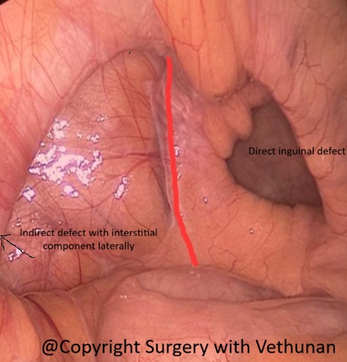
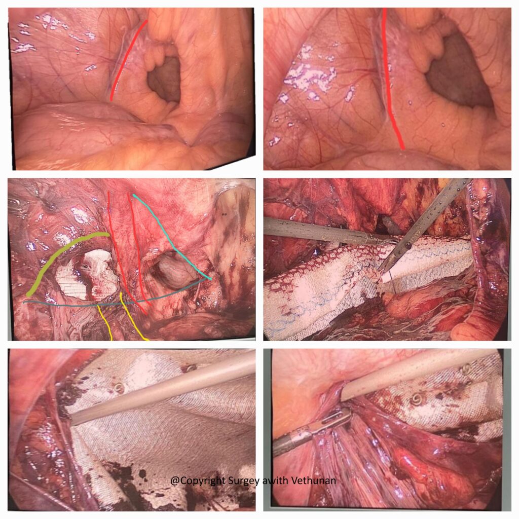
Laparoscopic TAPP exploration confirmed:
- A large direct inguinal hernia
- A laterally extending interstitial hernia dissecting between the abdominal wall muscle layers
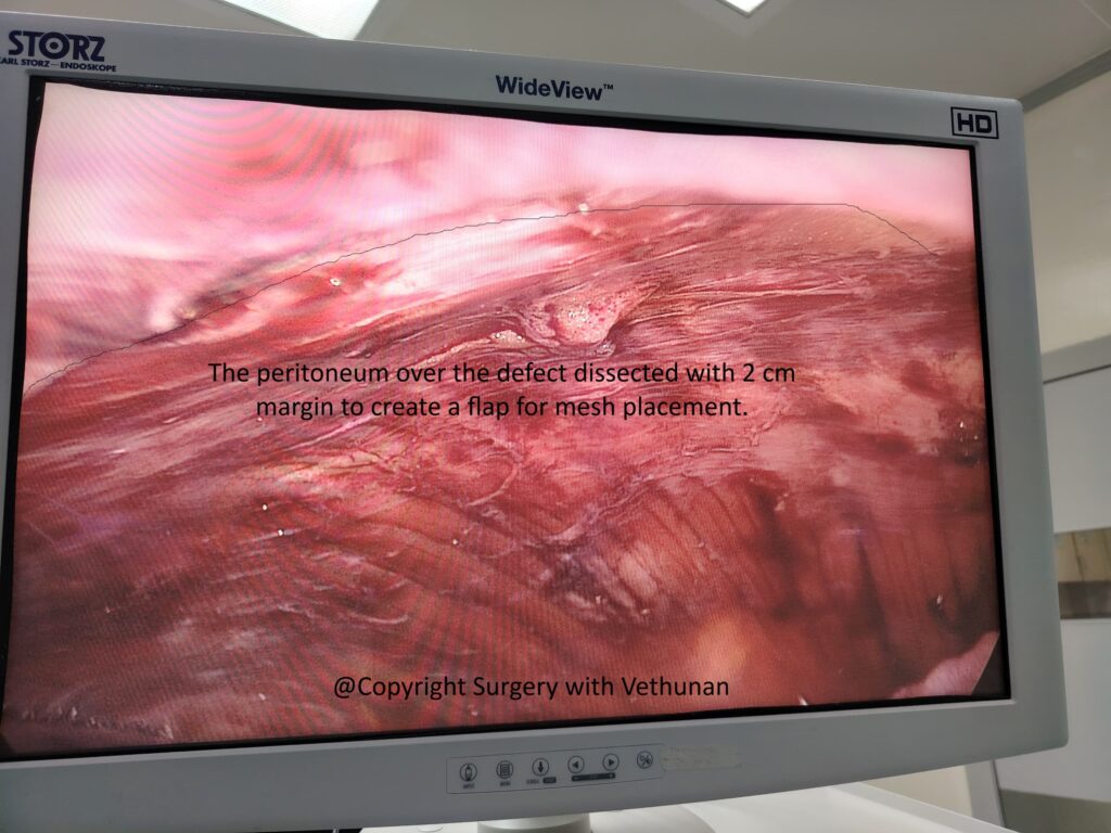
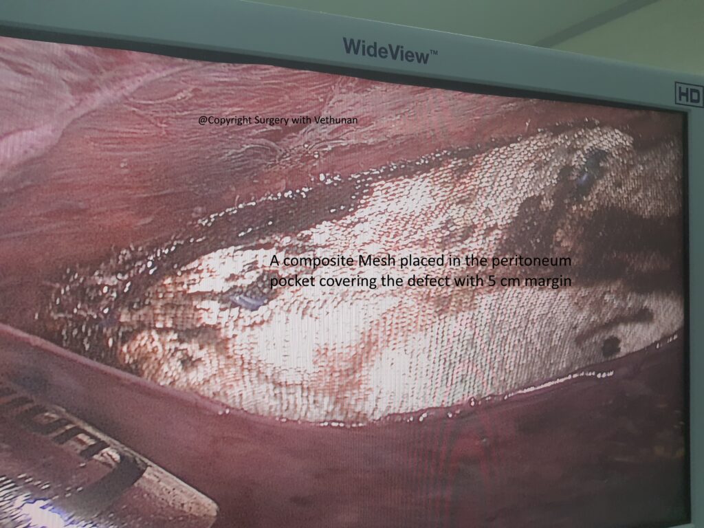
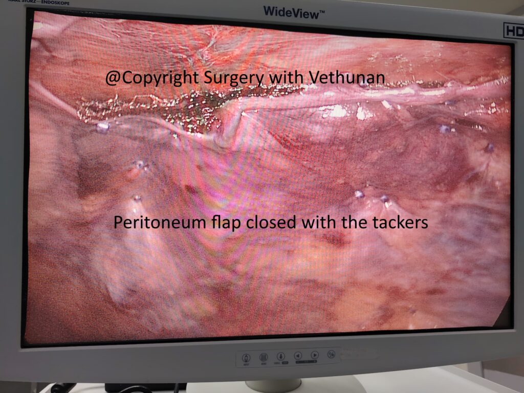
Following reduction of the hernia sac and dissection of the peritoneum flap, a large preperitoneal mesh was placed, covering the entire myopectineal orifice and the interstitial extension.
Case 2
Patient Presentation
A 45-year-old gentleman presented with a two-week history of a painful left lumbar swelling following minor blunt trauma.
He had no significant medical illnesses or previous abdominal surgery.
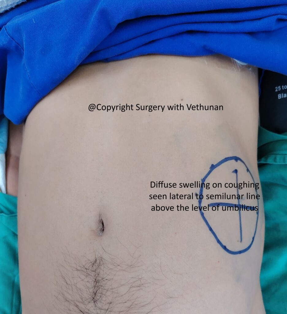
Clinical examination demonstrated a poorly defined swelling above the umbilicus and lateral to the semilunar line. The swelling demonstrated a positive cough impulse, suggesting an abdominal wall hernia despite its unusual location.
Because of the atypical presentation, the initial differential diagnosis included:
- Spigelian hernia
- Lumbar hernia
- Traumatic abdominal wall hernia
- Abdominal wall haematoma
Diagnostic Evaluation
Routine blood investigations were normal.
Given the uncertainty regarding the diagnosis and the patient’s persistent symptoms, laparoscopic exploration was undertaken.
Operative Findings
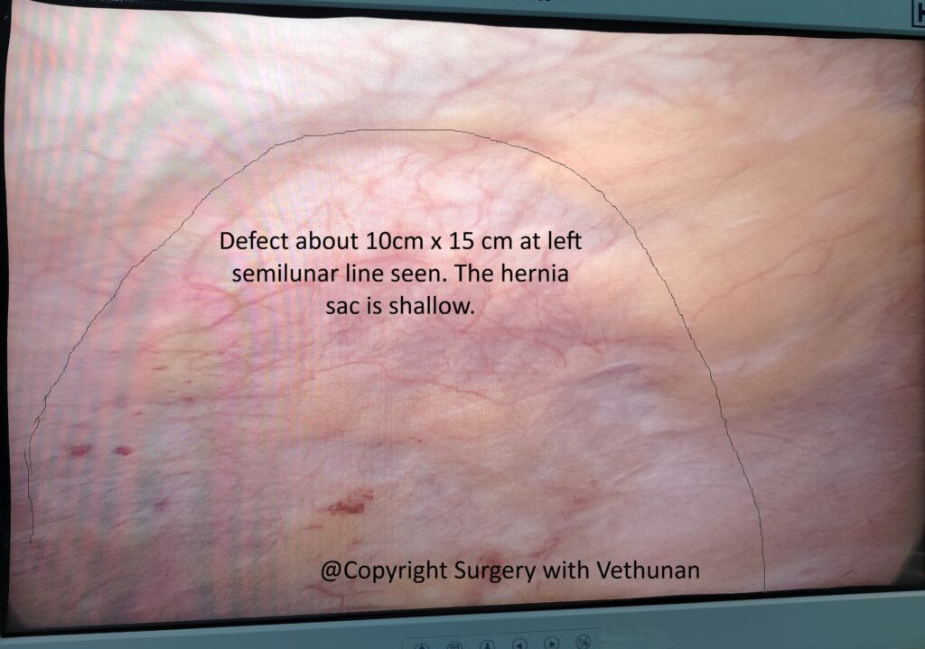
Laparoscopic TAPP exploration demonstrated an interstitial hernia at left lumbar area lateral to semilunar line and above umbilicus, with the hernia sac dissecting between the abdominal wall muscle layers. The hernia sac was shallow as it was an acute hernia.
The a peritoneum flap was dissected, and a large preperitoneal mesh was placed to reinforce the entire defect.
The procedure was completed laparoscopically without complication.
Discussion
Interstitial (interparietal) hernias are one of the rarest forms of abdominal wall hernia. Instead of protruding through the superficial fascia, the hernia sac tracks between the muscular and fascial layers of the abdominal wall. This unusual anatomical pathway often produces atypical clinical findings and contributes to delayed or missed diagnosis.
The two cases presented here demonstrate the wide spectrum of clinical presentations associated with this rare condition.
The first patient presented with a giant diffuse groin swelling that extended laterally towards the ASIS. Preoperative CT successfully identified both the direct inguinal defect and the interstitial extension, allowing accurate operative planning.
In contrast, the second patient presented with an acute painful lumbar swelling after minor trauma. The unusual location mimicked several other abdominal wall conditions, and the definitive diagnosis was only established during laparoscopic exploration.
These cases highlight that interstitial hernias should be considered in patients presenting with:
- Diffuse or atypical groin swelling
- Lateral abdominal wall swelling
- Chronic groin pain without an obvious hernia
- Positive cough impulse despite an unusual swelling location
- Persistent symptoms unexplained by conventional imaging or examination
Cross-sectional imaging, particularly CT scanning, is valuable for defining complex anatomy and identifying unusual hernia extensions. However, small or occult interstitial hernias may still be missed.
Laparoscopic TAPP repair offers significant advantages over conventional open surgery. It provides excellent visualisation of the entire myopectineal orifice, enables identification of occult or multiple defects, facilitates wide preperitoneal mesh placement, and avoids large incisions. In addition to being therapeutic, laparoscopy also serves as an important diagnostic tool in patients with atypical abdominal wall swellings.
Why This Case Series Is Unique
This series demonstrates two uncommon and distinctly different presentations of interstitial hernia successfully managed using the same minimally invasive technique.
Highlights include:
- Two rare cases of interstitial (interparietal) hernia
- Completely different clinical presentations despite the same underlying pathology
- One patient presenting as a giant diffuse groin swelling
- One patient presenting as a painful lumbar abdominal wall swelling following minor trauma
- Successful diagnosis using CT imaging in one patient and laparoscopic exploration in the other
- Definitive treatment with laparoscopic TAPP mesh repair in both cases
These cases reinforce the versatility of laparoscopic surgery in diagnosing and treating rare abdominal wall hernias.
Key Takeaways
- Interstitial (interparietal) hernias are rare and frequently underdiagnosed because of their atypical anatomy.
- Patients may present with diffuse groin swelling, lumbar swelling, chronic groin pain, or other unusual abdominal wall masses.
- CT imaging is useful for defining the anatomy of complex hernias but may not detect every case.
- Laparoscopic TAPP repair is both diagnostic and therapeutic, providing excellent exposure of the preperitoneal space while allowing comprehensive mesh reinforcement.
- Early recognition and appropriate minimally invasive management can achieve excellent clinical outcomes and minimise recurrence.
Both patients consented for publication of these cases