
Bouveret Syndrome: A Rare Cause of Gastric Outlet Obstruction
Bouveret syndrome is an exceptionally rare form of gallstone ileus, accounting for only 1–3% of all gallstone ileus cases. It occurs when a large gallstone erodes through the gallbladder wall into the duodenum via a cholecystoduodenal fistula and becomes impacted in the pylorus or proximal duodenum, resulting in gastric outlet obstruction.
First described by the French physician Léon Bouveret in 1896, the condition primarily affects elderly patients, with a reported median age of approximately 74 years. Women are affected more frequently than men due to the higher prevalence of gallstone disease.
Although uncommon, Bouveret syndrome is associated with significant morbidity because diagnosis is often delayed and patients frequently present with dehydration, malnutrition, and multiple medical comorbidities.
Patient Presentation
A 75-year-old woman presented with a one-month history of:
- Persistent vomiting
- Epigastric abdominal pain
- Poor oral intake
- Progressive weight loss
Clinical examination revealed mild dehydration and epigastric tenderness. Blood investigations demonstrated:
- Metabolic alkalosis
- Mild anaemia
- Leucocytosis
- Elevated urea consistent with dehydration
These findings are typical of prolonged gastric outlet obstruction.
Diagnostic Evaluation
Abdominal X-ray
Plain radiography demonstrated a radio-opaque lesion in the upper abdomen.
Although abdominal radiographs may reveal the classic Rigler’s triad (pneumobilia, bowel obstruction and an ectopic gallstone), this combination is seen in only a minority of patients.
Upper Gastrointestinal Endoscopy
Upper gastrointestinal endoscopy confirmed a large gallstone impacted within the second part of the duodenum.
Endoscopy is both diagnostic and therapeutic, allowing direct visualisation of the obstructing stone while providing an opportunity for endoscopic extraction or lithotripsy.
Multiple attempts at retrieval were unsuccessful because of the stone’s large size.
CT Scan
Contrast-enhanced CT demonstrated:
- Markedly dilated stomach and proximal duodenum
- Large impacted gallstone within D2
- Features consistent with Bouveret syndrome
CT is regarded as the imaging modality of choice, with reported diagnostic accuracy approaching 90% because it identifies the stone, level of obstruction, pneumobilia, and the biliary-enteric fistula.
Surgical Management
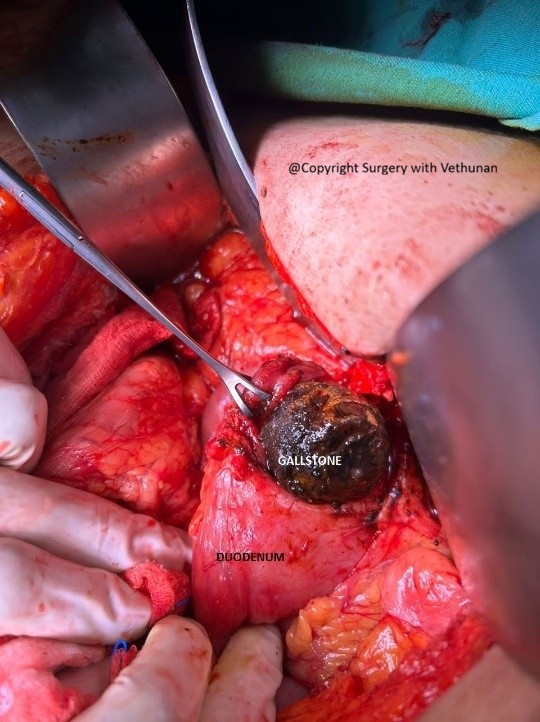
Open technique through a subcoastal incision.
Operative findings included:
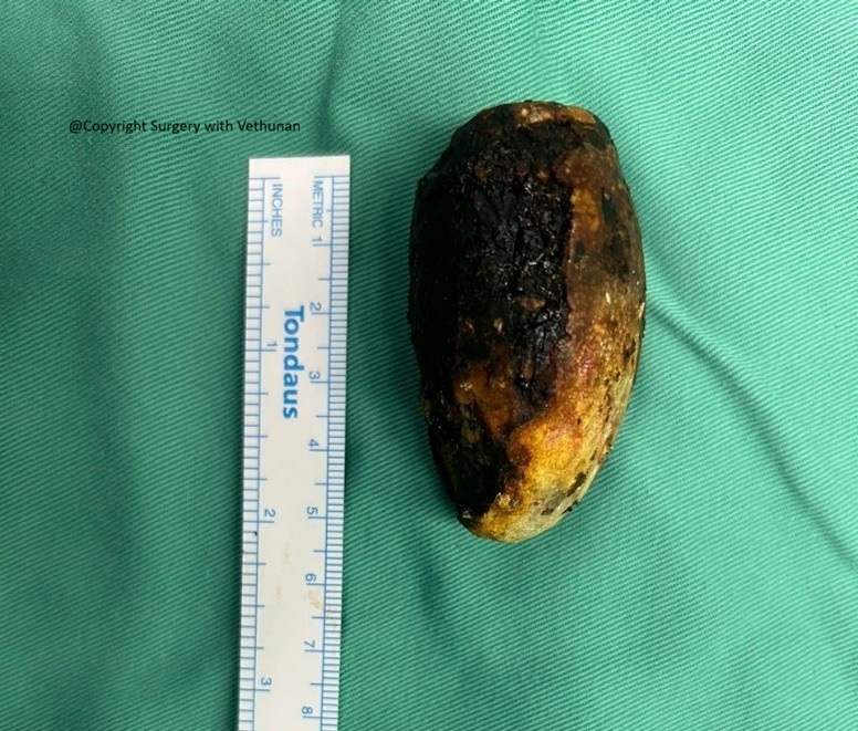
- Giant impacted gallstone measuring approximately 5 cm
- Dense inflammatory adhesions between the gallbladder and duodenum
- Cholecystoduodenal fistula
The following procedures were successfully performed:
- Duodenostomy
- Extraction of the impacted gallstone
- Cholecystectomy
- Excision of the cholecystoduodenal fistula
- Gastric decompression
- Repair of duodenostomy and fistula site with Vicryl 3/0
Understanding Bouveret Syndrome
Gallstones are common, affecting 10–20% of adults, yet fewer than 0.5% of patients with gallstones develop gallstone ileus, making Bouveret syndrome one of the rarest complications.
The disease develops gradually:
- Chronic inflammation causes the gallbladder to adhere to the adjacent duodenum.
- Pressure from a large gallstone leads to erosion and fistula formation.
- The gallstone migrates into the gastrointestinal tract.
- Instead of passing distally, the stone becomes lodged in the pylorus or proximal duodenum, causing gastric outlet obstruction.
Because symptoms evolve slowly over weeks, patients are often initially investigated for peptic ulcer disease, gastric cancer, or functional gastrointestinal disorders before the correct diagnosis is established.
Clinical Features
Patients commonly present with:
- Persistent nausea and vomiting
- Epigastric pain
- Early satiety
- Progressive weight loss
- Dehydration
- Electrolyte imbalance
Less commonly, patients may develop:
- Haematemesis
- Upper gastrointestinal bleeding
- Acute cholangitis
- Pancreatitis
Due to the non-specific nature of these symptoms, maintaining a high index of suspicion is essential, particularly in elderly patients with known gallstone disease.
Treatment Options
Management should be individualised based on the patient’s physiological status, stone size, and available expertise.
Initial Management
- Fluid resuscitation
- Electrolyte correction
- Nasogastric decompression
- Nutritional optimisation
- Antibiotics when infection is suspected
Endoscopic Treatment
Whenever feasible, endoscopic therapy is attempted first.
Available techniques include:
- Basket retrieval
- Mechanical lithotripsy
- Laser lithotripsy
- Electrohydraulic lithotripsy
However, reported endoscopic success rates remain relatively low (around 30–40%) because most stones are larger than 2.5 cm.
Surgical Treatment
Surgery remains the gold standard when:
- Endoscopic removal fails
- The stone is very large
- There is perforation or sepsis
- The patient develops ongoing obstruction
Depending on the patient’s condition, surgery may involve:
- Enterolithotomy or duodenostomy alone
- One-stage surgery with cholecystectomy and fistula repair
- Two-stage surgery in selected high-risk patients
Why This Case Is Unique
Several features make this case particularly noteworthy:
- Bouveret syndrome is an exceptionally rare surgical emergency.
- The patient had a giant 5 cm gallstone, significantly larger than most reported cases.
- Endoscopic retrieval was attempted before surgery, reflecting current evidence-based management.
- Definitive surgical treatment included both stone extraction and management of the biliary fistula.
- Such cases require close collaboration between surgeons, gastroenterologists, anaesthetists, and radiologists to achieve the best outcomes.
Key Takeaways
- Bouveret syndrome is a rare but important cause of gastric outlet obstruction.
- Elderly patients with persistent vomiting and a history of gallstones should prompt consideration of this diagnosis.
- Contrast-enhanced CT and upper gastrointestinal endoscopy are the most useful diagnostic investigations.
- Endoscopic therapy should be attempted first when technically feasible.
- Surgery remains the definitive treatment for large impacted gallstones or when endoscopic treatment is unsuccessful.
- Early diagnosis and multidisciplinary management are essential for improving patient outcomes.
Consent for online publishing of image and case obtained from patient