Giant Omental Teratoma: A Rare Intra-Abdominal Tumour in a 35-Year-Old Woman
An omental teratoma is an exceptionally rare tumour arising within the omentum. Teratomas are tumours containing tissues derived from more than one embryonic germ cell layer and are more commonly encountered in the gonads, particularly the ovaries.
A teratoma arising primarily from the omentum is extremely uncommon and can present a significant diagnostic challenge because a large omental mass may resemble an ovarian tumour, mesenteric tumour, gastrointestinal stromal tumour, liposarcoma, or other intra-abdominal neoplasm.
.
Patient Presentation
A 35-year-old lady was evaluated for a large intra-abdominal mass. She was referred by a gynecologist.
Clinical assessment revealed a sizeable intrabdominal mass, which was mobile at umbilical region.
Patient had occasional pain at that site. Her gynecology history was normal. She had 2 previous C-section. Tumour makers taken was normal.
CT Imaging
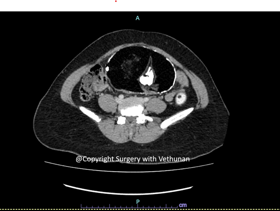
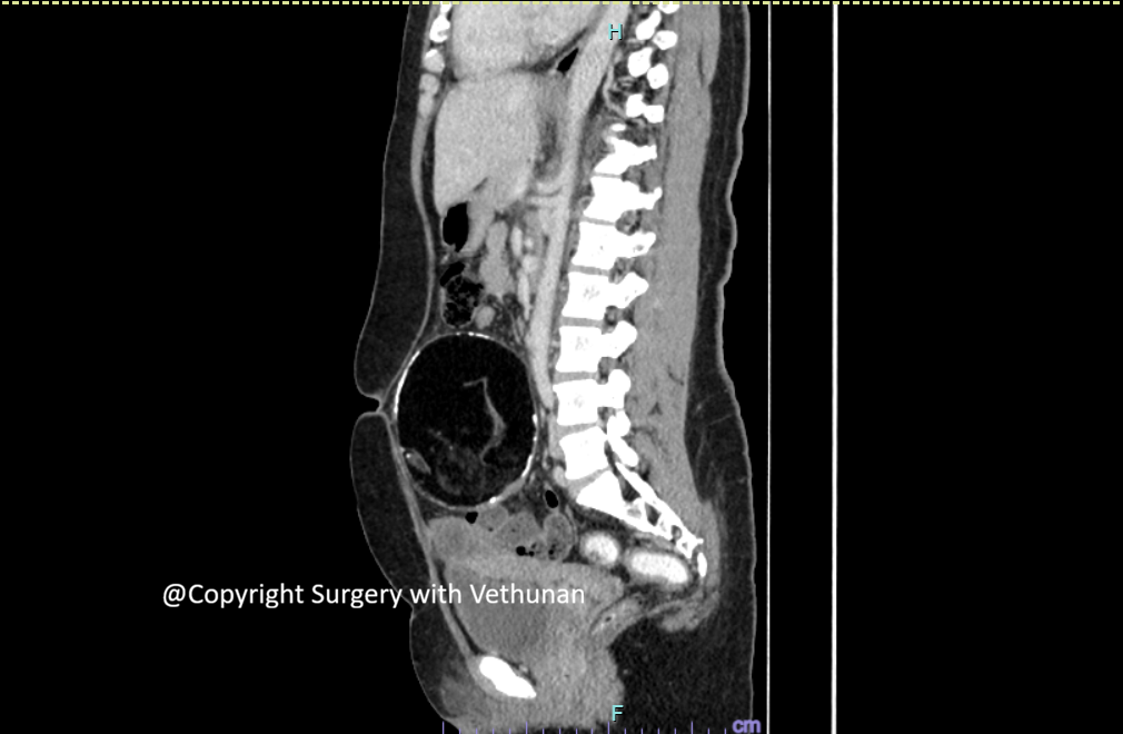
CT imaging demonstrated a large intra-abdominal mass measuring approximately 15 × 14 cm.
The lesion demonstrated features suggestive of a teratomatous tumour, with the presence of different tissue components within the mass.
An important part of the radiological assessment was establishing that the lesion was separate from the ovaries and other major intra-abdominal organs, supporting an omental origin.
Surgical Management
Because of the large size of the tumour and uncertainty regarding its exact origin, the patient underwent exploratory laparotomy.
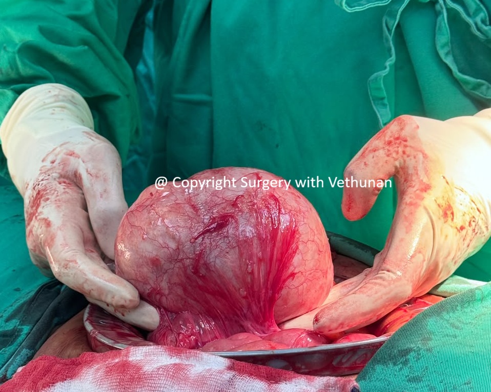
At laparotomy, a large tumour measuring approximately 15 × 14 cm was identified arising from the omentum.
The tumour was carefully mobilised from the surrounding structures.
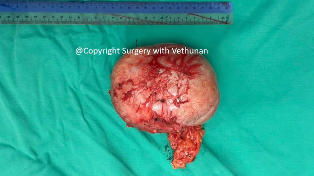
Complete surgical excision was performed with preservation of the adjacent abdominal organs.
Teratoma with pedicle from omentum and attachment to the base of mesentery
Excised teratoma with pedicle
Histopathology
Teratomas contain tissues derived from different germ cell layers and may contain mature tissues such as:
Adipose tissue
Cartilage
Bone
Hair
The histological examination determined that the lesion is mature and benign.
In this case, the final diagnosis was omental teratoma.
Understanding Omental Teratoma
Teratomas most commonly occur within the gonads. Extragonadal teratomas are much less common and can occur in locations such as the mediastinum, retroperitoneum and, rarely, the mesentery or omentum.
The exact mechanism by which a teratoma develops in the omentum remains uncertain. They are typically found in women of reproductive age, but may also appear in young girls and in older women. Several theories have been proposed, including:
Primary teratomas of the omentum may originate from displaced germ cells.
Teratomas may develop in a supernumerary ovary of the omentum.
Teratomas may result from autoamputation of an ovarian dermoid cyst with secondary implantation into the greater omentum.
Because of their rarity, establishing that the tumour is truly primary to the omentum requires careful radiological, operative and pathological assessment to exclude a gonadal or other primary tumour.
Mature teratomas are generally benign and have an excellent prognosis following complete excision. Laparoscopy should be the first choice when possible, unlike this case (due to its size).
Immature or malignant teratomas require more extensive oncological assessment and may require adjuvant chemotherapy depending on the pathological findings.
Why This Case Is Unique
This case is particularly noteworthy because:
The tumour arose from the omentum, an exceptionally unusual location for a teratoma.
The lesion was large, measuring 15 × 14 cm.
Establishing the organ of origin was important in distinguishing it from more common ovarian and retroperitoneal tumours.
Key Takeaways
Omental teratoma is an extremely rare extragonadal teratoma.
Large omental tumours can mimic ovarian, mesenteric, retroperitoneal and other intra-abdominal tumours.
CT imaging is important for defining the tumour’s size, composition and relationship to surrounding organs.
Establishing that the ovaries and other potential primary sites are normal is important when diagnosing a primary omental teratoma.
Complete surgical excision is the mainstay of treatment for a resectable lesion.
Histopathology is essential to distinguish a benign mature teratoma from immature or malignant variants.
Consent for publication and picture obtained from patient.