
Giant Left Adrenal Myelolipoma Causing Compression of the Left Renal Vessels
Adrenal myelolipoma is a rare, benign tumour composed of mature fat and normal bone marrow elements. Although it accounts for approximately 3–5% of adrenal incidentalomas, most lesions are small, asymptomatic, and discovered incidentally during imaging performed for unrelated conditions.
Large or “giant” adrenal myelolipomas (typically larger than 10 cm) are uncommon. As they enlarge, they may compress surrounding organs and blood vessels, leading to abdominal pain or a palpable abdominal mass. Surgical excision is recommended for symptomatic tumours, large lesions, or when malignancy cannot be confidently excluded.
Patient Presentation
A 45-year-old woman presented with a six-month history of persistent upper abdominal pain.
Her medical history was significant for paraplegia following traumatic spinal cord injury at the age of 20 years. Despite her neurological disability, she had remained medically stable for many years before developing progressive upper abdominal discomfort.
Clinical examination revealed:
- A large, firm mass occupying the left hypochondrium
- The mass was non-mobile and deep seated
- No features of acute abdomen or bowel obstruction were present
Given the location of the pain and the palpable mass, further investigation was undertaken.
Diagnostic Evaluation
Laboratory Investigations
Routine blood investigations were performed as part of the initial assessment.
Results demonstrated:
- Normal full blood count
- Normal renal function
- Normal liver function tests
- Normal inflammatory markers
- Normal serum electrolytes
The absence of biochemical abnormalities suggested that the patient’s symptoms were related to the mass effect of the tumour rather than an inflammatory or systemic process.
Upper Gastrointestinal Endoscopy (OGDS)
Upper gastrointestinal endoscopy was performed to investigate the patient’s upper abdominal pain.
The examination was entirely normal, with no evidence of:
- Peptic ulcer disease
- Gastric outlet obstruction
- Gastric or duodenal malignancy
- Upper gastrointestinal bleeding
As no intraluminal pathology was identified, cross-sectional imaging was pursued to evaluate the palpable abdominal mass.
Contrast-Enhanced CT Scan
Contrast-enhanced CT of the abdomen demonstrated:
- A large (15cm diameter), well-circumscribed left retroperitoneal mass arising from the left adrenal gland
- Significant compression of the left renal artery and renal vein, without radiological evidence of vascular invasion
- Abundant macroscopic fat interspersed with soft tissue elements, producing the characteristic appearance of an adrenal myelolipoma
- Well-defined tumour margins with preservation of the surrounding tissue planes
- Tumour abuts the pancrease and splenic vein
- No enlarged regional lymph nodes or distant metastatic disease
The imaging features strongly favoured a diagnosis of giant left adrenal myelolipoma, while also demonstrating the technical complexity of surgical resection because of the tumour’s close relationship to the renal hilum and major retroperitoneal vessels.
Endocrine (Functional) Assessment
As recommended for all adrenal incidentalomas, a comprehensive hormonal evaluation was undertaken to exclude a functioning adrenal tumour.
The biochemical work-up demonstrated normal adrenal function, including:
- Normal plasma or urinary metanephrines, excluding pheochromocytoma
- Normal overnight dexamethasone suppression test, excluding autonomous cortisol secretion (Cushing syndrome)
- Normal aldosterone-renin ratio where clinically indicated, excluding primary hyperaldosteronism
These findings confirmed that the lesion was a non-functioning adrenal tumour, consistent with the typical behaviour of adrenal myelolipoma.
Surgical Management
The primary objectives of surgery were:
- Complete removal of the adrenal tumour
- Preservation of the left kidney and renal vessels
- Protection of surrounding retroperitoneal structures
- Histopathological confirmation of the diagnosis
Access: Midline Laparotomy
Meticulous dissection was done keeping with close proximity to the renal hilum, pancreas, spleen, diaphragm, and major abdominal vessels.
- The lesser sac was opened
- Transverse colon and splenic flexure was mobilzed inferomedially
- Posterior wall of lesser sac incised below the inferior margin of pancrease
- Preservation of splenic vein with meticulous disection
- Pancrease with spleen was flipped superomedially
- The inferior border of tumour was dissected away from the stretched left renal vessel
- The kidney was separated from tumour by dissecting along the fascia septum separating it from left kidney
- The tumour was then freed from posterior wall after dividing attaching ligaments to diaphragm
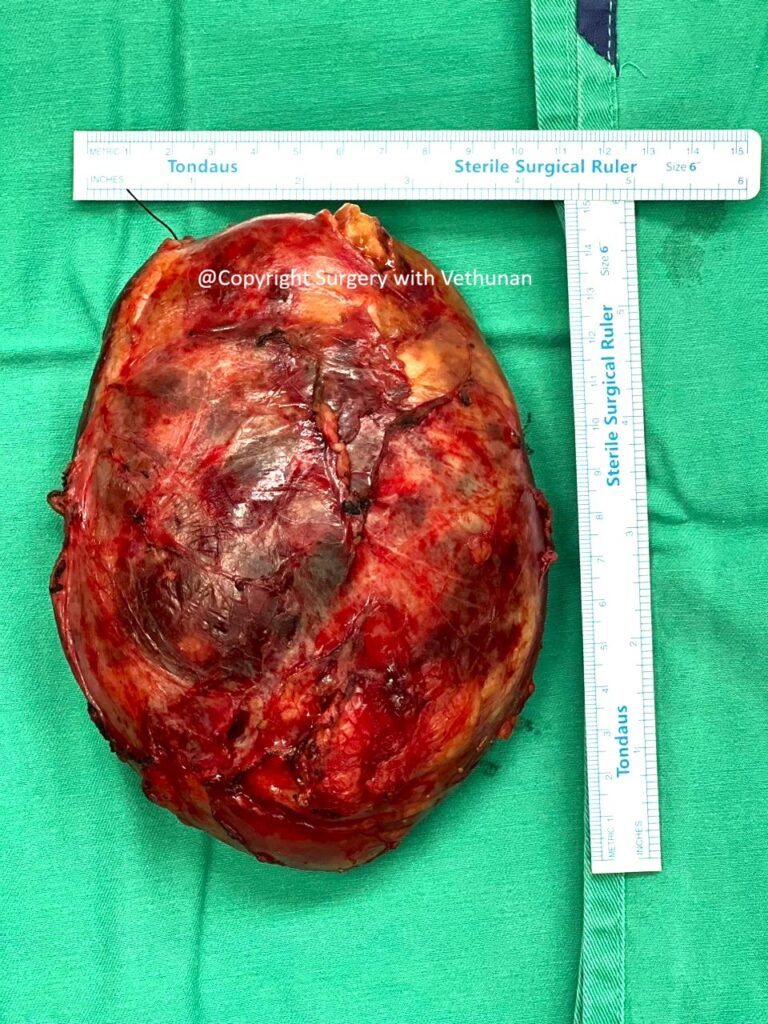
Histopathological Examination
Histopathological examination confirmed the diagnosis of adrenal myelolipoma.
Microscopic analysis demonstrated:
- Mature adipose tissue
- Trilineage haematopoietic elements
- No evidence of malignancy
These findings confirmed the benign nature of the tumour.
Understanding Adrenal Myelolipoma
Adrenal myelolipoma is a rare benign tumour of the adrenal gland. The exact cause remains uncertain, although several theories have been proposed, including metaplasia of adrenal cortical cells in response to chronic stress or inflammation.
Most patients are diagnosed between the fifth and seventh decades of life, and there is no strong gender predominance.
The widespread use of CT and MRI has led to increasing detection of adrenal incidentalomas, with myelolipoma now recognised more frequently than in previous decades.
Although usually asymptomatic, giant tumours may cause symptoms due to compression of adjacent organs, including:
- Persistent abdominal or flank pain
- Early satiety
- Palpable abdominal mass
- Compression of the kidney or renal vessels
- Rare spontaneous retroperitoneal haemorrhage
Treatment Options
Management depends on tumour size, symptoms, imaging characteristics, and hormonal function.
Observation
Small (<4–5 cm), asymptomatic, non-functioning lesions with characteristic imaging features may be monitored with periodic imaging.
Surgical Excision
Surgery is recommended when:
- The tumour causes symptoms
- The lesion is large (generally >6–7 cm)
- There is uncertainty regarding the diagnosis
- There is significant growth during follow-up
- There is concern for spontaneous haemorrhage
Complete surgical excision is usually curative.
Why This Case Is Unique
Several features make this case particularly noteworthy:
- A rare giant left adrenal myelolipoma presenting with chronic upper abdominal pain.
- Significant compression of the left renal vessels, creating additional surgical complexity.
- Successful exclusion of hormonally active adrenal disease through comprehensive endocrine assessment.
- Surgical management in a patient with long-standing traumatic paraplegia, requiring careful perioperative planning and multidisciplinary care.
- Complete tumour excision with histopathological confirmation of a benign adrenal myelolipoma.
Key Takeaways
- Adrenal myelolipoma is a rare benign tumour composed of mature fat and bone marrow tissue.
- Most lesions are discovered incidentally, but large tumours may present with abdominal pain or a palpable mass due to compression of surrounding structures.
- CT imaging and endocrine evaluation are essential components of assessment.
- Surgical excision is the treatment of choice for symptomatic or giant lesions.
- Complete resection offers an excellent prognosis, with recurrence being exceptionally rare.
Consent for publication obtained from patient