Duodenojejunal Junction Adenocarcinoma Presenting with Intestinal Obstruction: Treated with Segmental Resection
Adenocarcinoma of the duodenojejunal junction (DJ junction) is an exceptionally uncommon gastrointestinal malignancy. Primary small bowel adenocarcinoma accounts for only a small proportion of gastrointestinal cancers, and tumours arising specifically at the duodenojejunal junction are particularly rare.
Because of its unusual location and non-specific symptoms, diagnosis can be challenging. Patients may initially present with abdominal pain, vomiting, weight loss, anaemia, or symptoms of intestinal obstruction.
Patient Presentation
A 45-year-old lady presented with symptoms of intestinal obstruction.
She experienced:
- Progressive abdominal distension
- Recurrent vomiting
- Abdominal pain
- Inability to tolerate oral intake
- Constipation
The relatively young age and absence of previous abdominal surgery made a malignant cause of obstruction less immediately apparent.
Clinical examination was consistent with intestinal obstruction, prompting further investigation.
Diagnostic Evaluation
Laboratory Investigations
Initial blood investigations were performed to assess the severity of the obstruction and to prepare the patient for possible surgery.
The laboratory findings showed systemic inflamation with hyponatremia.
Imaging
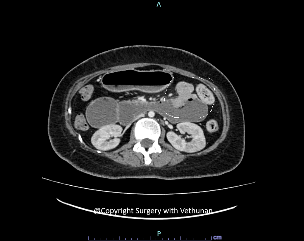
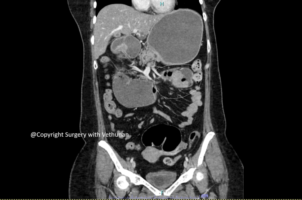
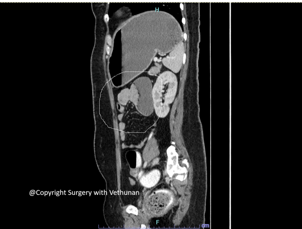
CT of the abdomen and pelvis demonstrated:
- Dilated proximal duodenum and stomach
- A transition point at the duodenojejunal junction
- An irregular circumferential lesion at the DJ junction
- No obvious distant metastatic disease on initial imaging
Endoscopic Evaluation
Surgical Management
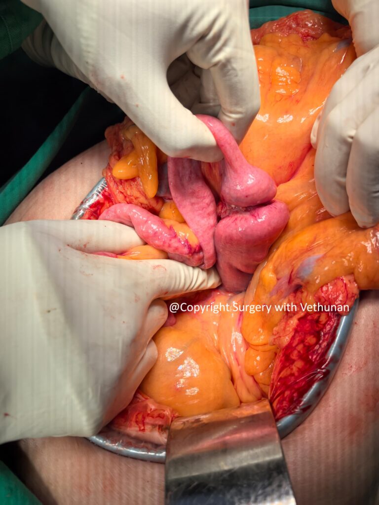
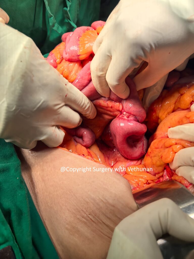
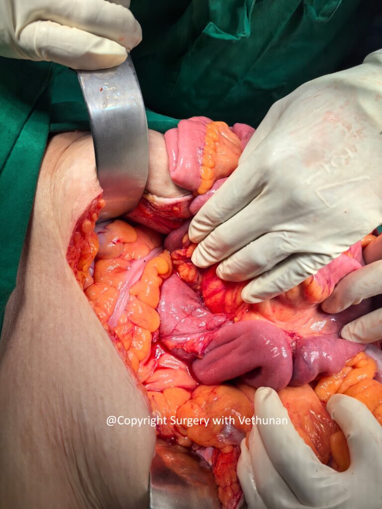
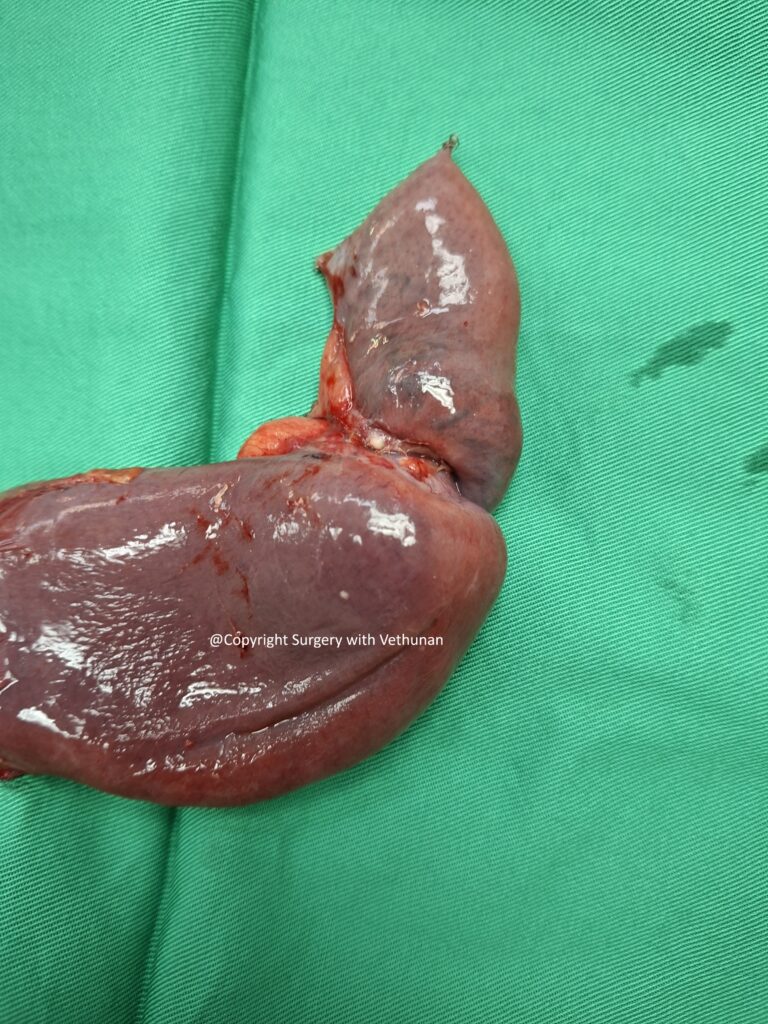
At surgery, an annular tumour was identified at the duodenojejunal junction, causing significant luminal narrowing and proximal bowel dilatation.
The tumour had no local invasion and was free from SMA and SMV. There was no peritoneal nodules or liver metastasis. The rest of bowel was tumour free.
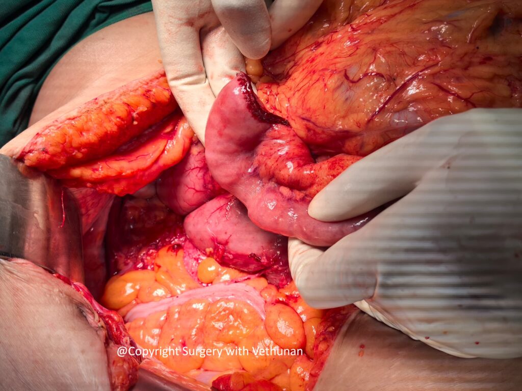
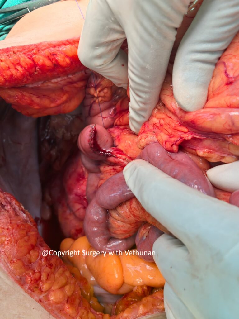
The tumour was resected with appropriate margins together with the associated mesentery and regional lymph nodes.
Due to it difficult anatomy a full Kocher’s maneuver was taken to mobilize the duodenum and the Ligament of Treitz was divided. The entire small bowel mesentery was mobilized and flipped superiorly after a Cattle -Braasch maneuver.
Following tumour resection, gastrointestinal continuity was restored with a side to side stapled jejunum- duodenum (D3) anastomosis.
Histopathological Examination
Duodenal adenocarcinoma moderately differentiated with lymphovascular invasion. pT4 pN0
Understanding Small Bowel Adenocarcinoma
Small bowel adenocarcinoma is uncommon compared with colorectal, gastric and pancreatic cancers.
The rarity of the disease contributes to diagnostic delay because symptoms such as abdominal pain, nausea, vomiting and weight loss are non-specific.
Risk factors and associated conditions include:
- Crohn’s disease
- Coeliac disease
- Familial adenomatous polyposis
- Lynch syndrome
- Peutz–Jeghers syndrome
- Other hereditary cancer syndromes
However, many patients have no identifiable predisposing condition.
The duodenum is the most frequent site of small bowel adenocarcinoma, while tumours arising specifically at the duodenojejunal junction are considerably less common.
Surgery
For resectable, localised small bowel adenocarcinoma, complete surgical resection with negative margins and appropriate regional lymphadenectomy remains the cornerstone of treatment.
The extent of surgery depends on tumour location.
For tumours near the duodenojejunal junction, the surgeon must carefully balance:
- Adequate oncological margins (5cm distal and proximal margin)
- Regional lymph node clearance (minimum 8, adequate lymph node clearance is important for prognosis)
- Preservation of pancreatic and mesenteric structures
- Restoration of gastrointestinal continuity
The anatomy of the DJ junction can make surgical planning particularly challenging because of its proximity to the pancreas, superior mesenteric vessels, transverse mesocolon and retroperitoneal structures.
The role of chemotherapy depends on the final pathological stage and multidisciplinary assessment.
Adjuvant chemotherapy is generally considered for patients with:
- Node-positive disease
- High-risk pathological features
- Selected high-risk stage II disease
For patients with MSI-high/dMMR tumours, the potential role of immunotherapy should also be considered, particularly in advanced disease.
Prognosis
The prognosis of SBA remains poor even after surgical resection. The 5-year survival ranges from 20%-50%. Prognosis is strongly influenced by:
- Stage at diagnosis(The stage-wise 5-year OS is 55%, 50%, 30%, and 5% for Stages I, II, III, and IV, respectively)
- Lymph node involvement( Inadequate lymph node clearance cannot be compensated with adjuvant chemotherapy)
- Completeness of tumour resection
- Tumour differentiation
- Lymphovascular and perineural invasion
- Presence of metastatic disease
Unfortunately, the rarity of small bowel adenocarcinoma means that many patients are diagnosed only after the tumour has produced significant symptoms or complications such as obstruction.
Why This Case Is Unique
his case is particularly noteworthy because:
- The patient was relatively young at 45 years of age.
- The tumour arose at the duodenojejunal junction, an uncommon location for gastrointestinal adenocarcinoma.
- The initial presentation was mechanical intestinal obstruction, rather than the more gradual symptoms of abdominal discomfort or weight loss.
- The unusual location presents significant diagnostic and surgical challenges.
- Definitive surgical resection provides both relief of obstruction and oncological treatment.
Key Takeaways
- Duodenojejunal junction adenocarcinoma is extremely rare.
- Small bowel adenocarcinoma can present with vague symptoms and may remain undiagnosed until complications develop.
- Mechanical obstruction in an otherwise unexplained patient should prompt consideration of an obstructing tumour.
- CT is an important investigation for defining the site of obstruction, tumour extent and metastatic disease.
- Complete surgical resection with appropriate lymphadenectomy is the mainstay of treatment for resectable disease.
- Histopathological staging and molecular testing, including MMR/MSI assessment, help guide postoperative management.
- Multidisciplinary management is important because of the rarity and complex anatomy of these tumours.
Consent for online publishing of image and case obtained from patient